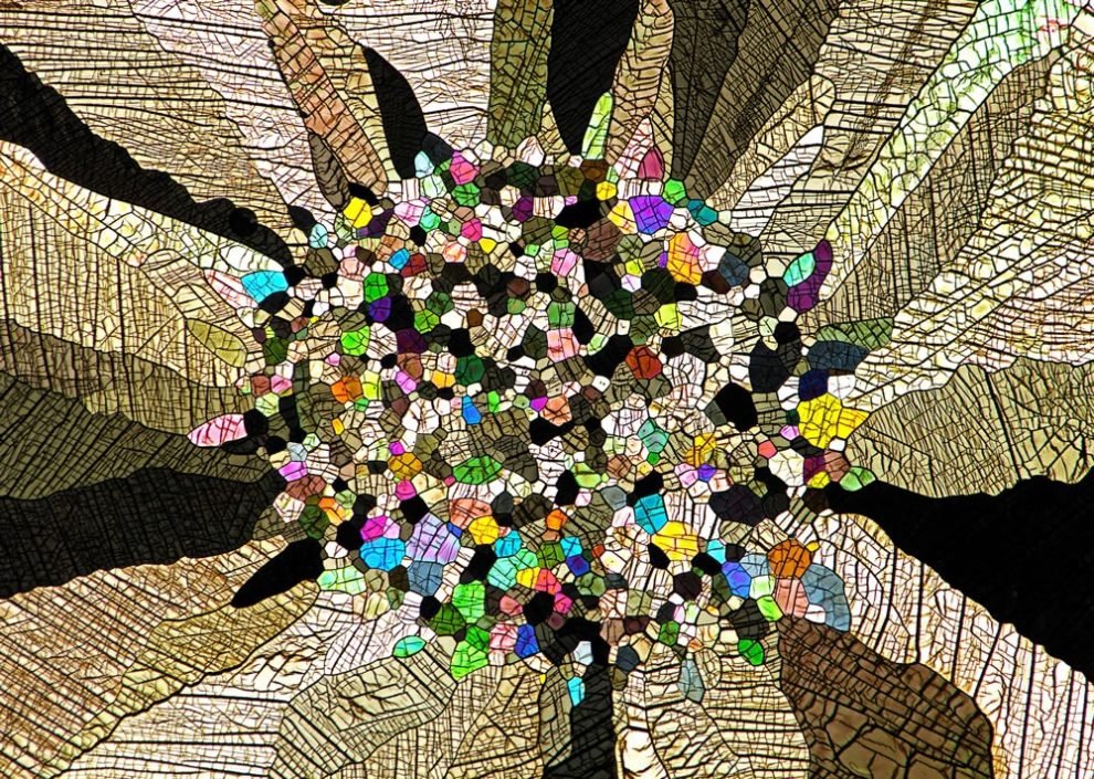
Rodent optic nerve head showing astrocytes (yellow), contractile proteins (red) and retinal vasculature (green) by Hassanain Qambari & Jayden Dickson
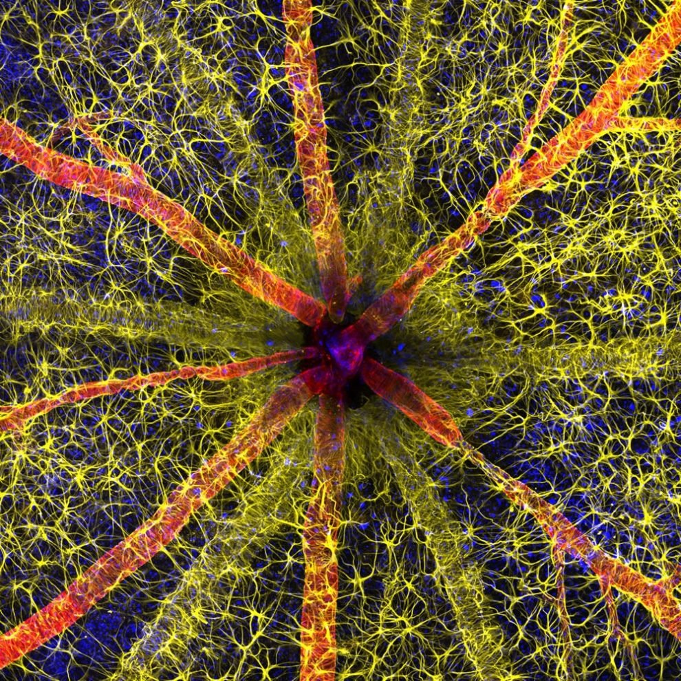
The Photomicrography Competition has recently concluded, leaving us in awe of the magnificent champions that have emerged. One of the most notable winners is Hassanain Qambari, who claimed the prestigious crown for his captivating portrayal of a rodent’s optic nerve head. Qambari’s image not only captured the anatomical intricacies of the rodent’s eye but also seemed to possess a vibrant energy that brought it to life before our very eyes.
This esteemed competition, born in the creative cauldron of 1975, was established with a noble purpose – to pay homage to the pioneers of light microscope photography. Since its inception, the Nikon Small World Competition has evolved into a grand spectacle, acting as a curator for the breathtaking masterpieces produced by photomicrographers from a diverse range of scientific fields. It has become a revered theater where these talented individuals can showcase their artistry and provide us with glimpses into the magnificent world that exists at a microscopic level.
This year’s competition was especially momentous, as it attracted an overwhelming number of participants from all corners of the globe. A staggering total of nearly 1,900 captivating snapshots poured in, originating from 72 different nations. This flood of submissions served as a testament to the ever-growing fascination and appreciation for the beauty that lies hidden within the tiniest of organisms and structures. The competition, however, is more than just a mere contest. It transforms into a global carnival of microscopic artistry, where the invisible entities of our world are brought to life in the most vivid and awe-inspiring ways.
The photographers who participate in this event possess a unique ability to explore the hidden realms that lie beyond the naked eye. They peel back the layers of reality, revealing the intricate beauty that often goes unnoticed but is undeniably present all around us. Each photograph submitted to the competition tells a story, a narrative of the unseen wonders that exist within our world. They bring to light the delicate intricacies of nature, showcasing the symphony of colors, patterns, and textures that reside within even the smallest of organisms.
These images serve as a reminder of the boundless creativity and complexity that permeates every corner of our existence. As we immerse ourselves in the captivating artistry of the winners and participants of the Photomicrography Competition, we are transported into a world that is both familiar and yet entirely foreign. It is a world where the mundane is transformed into the extraordinary and where the invisible becomes impossibly vivid. It is a testament to the power of science, technology, and human creativity to reveal the hidden wonders that lie just beyond our perception.
In the end, the Photomicrography Competition is not just a celebration of the champions who emerge victorious. It is a celebration of the human spirit, the relentless curiosity that drives us to explore and uncover the mysteries of the universe. It is a reminder that there is beauty to be found in even the smallest and most hidden of places, and that our world is a canvas for endless discovery and wonder.
More: Nikon Small World, Instagram
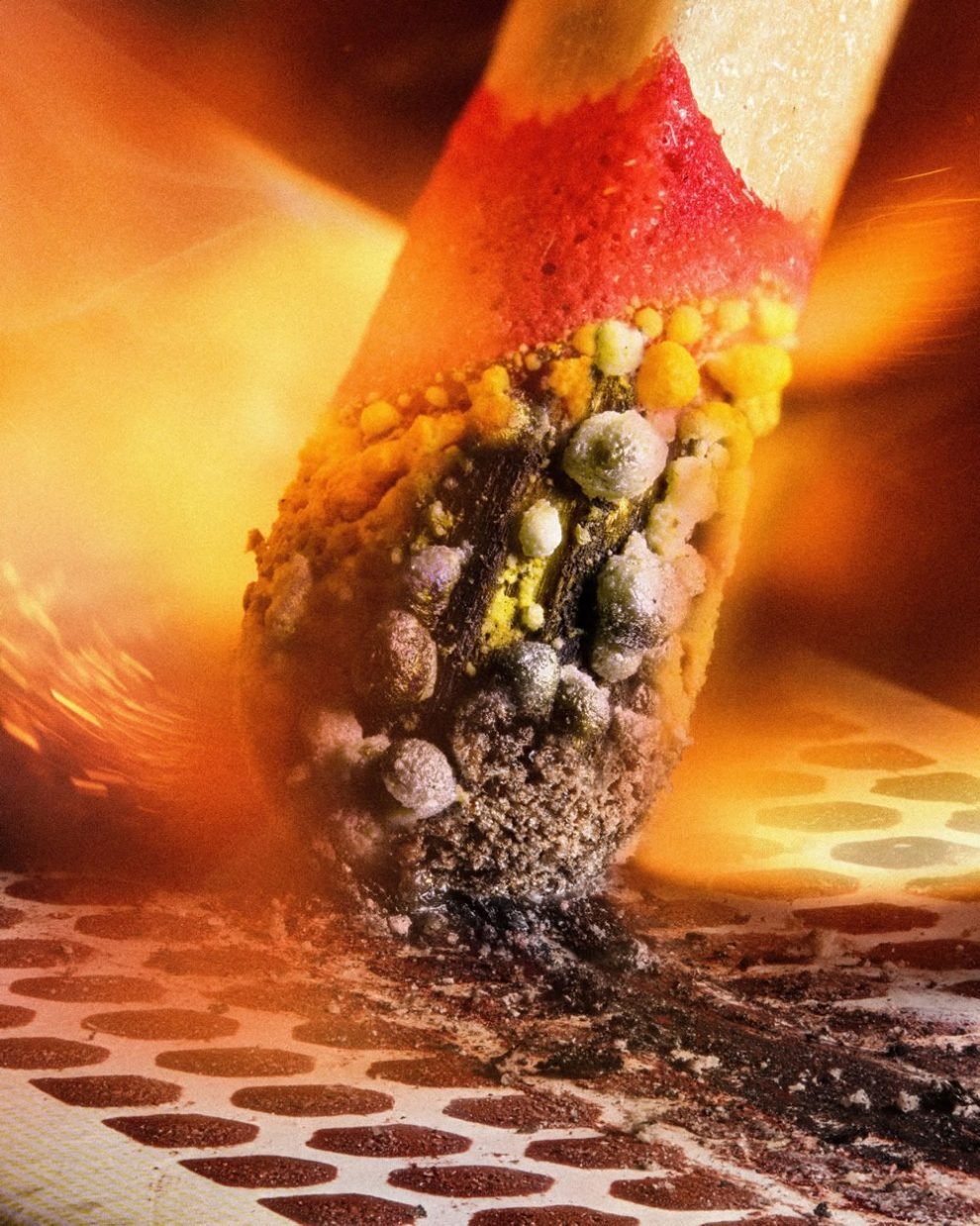
Matchstick igniting by the friction surface of the box by Ole Bielfeldt
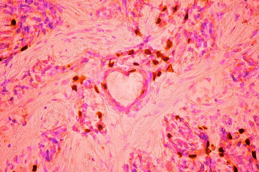
Breast cancer cells by Malgorzata Lisowska
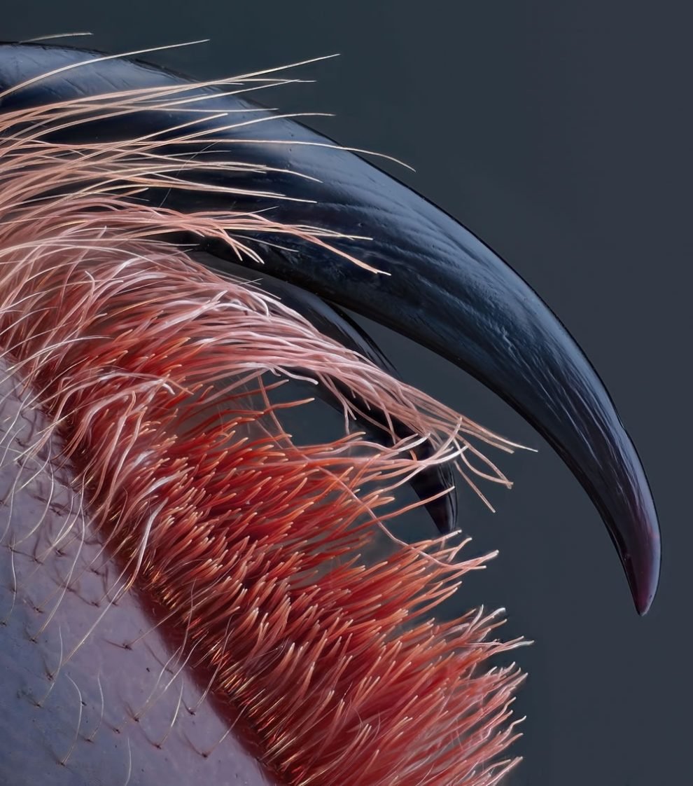
Venomous fangs of a small tarantula by John-Oliver Dum
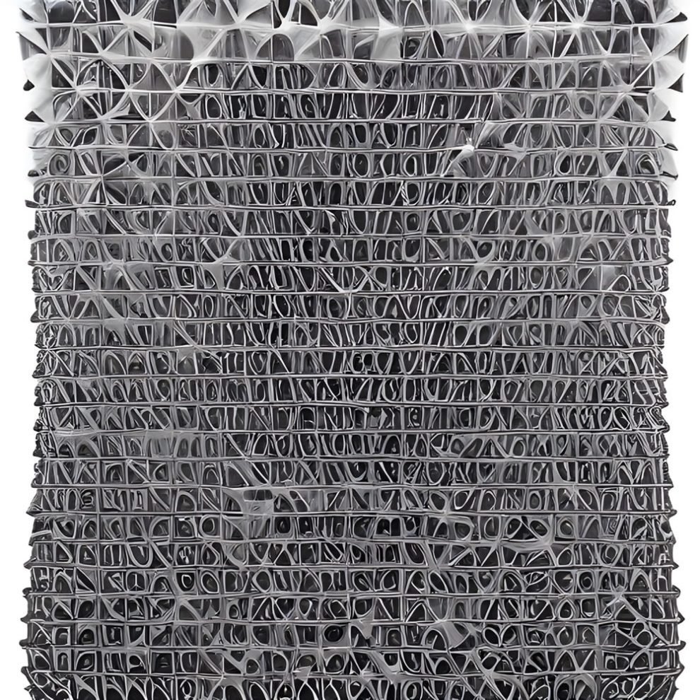
Auto-fluorescing defensive hairs covering the leaf surface of Eleagnus angustifolia exposed to UV light by Dr. David Maitland
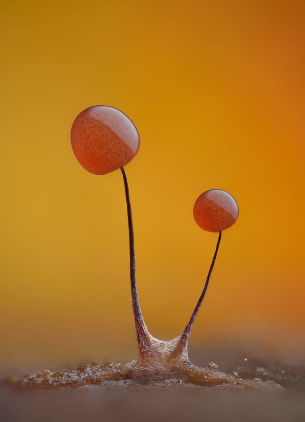
Slime mold (Comatricha nigra) showing capillitial fibers through its translucent peridium by Timothy Boomer
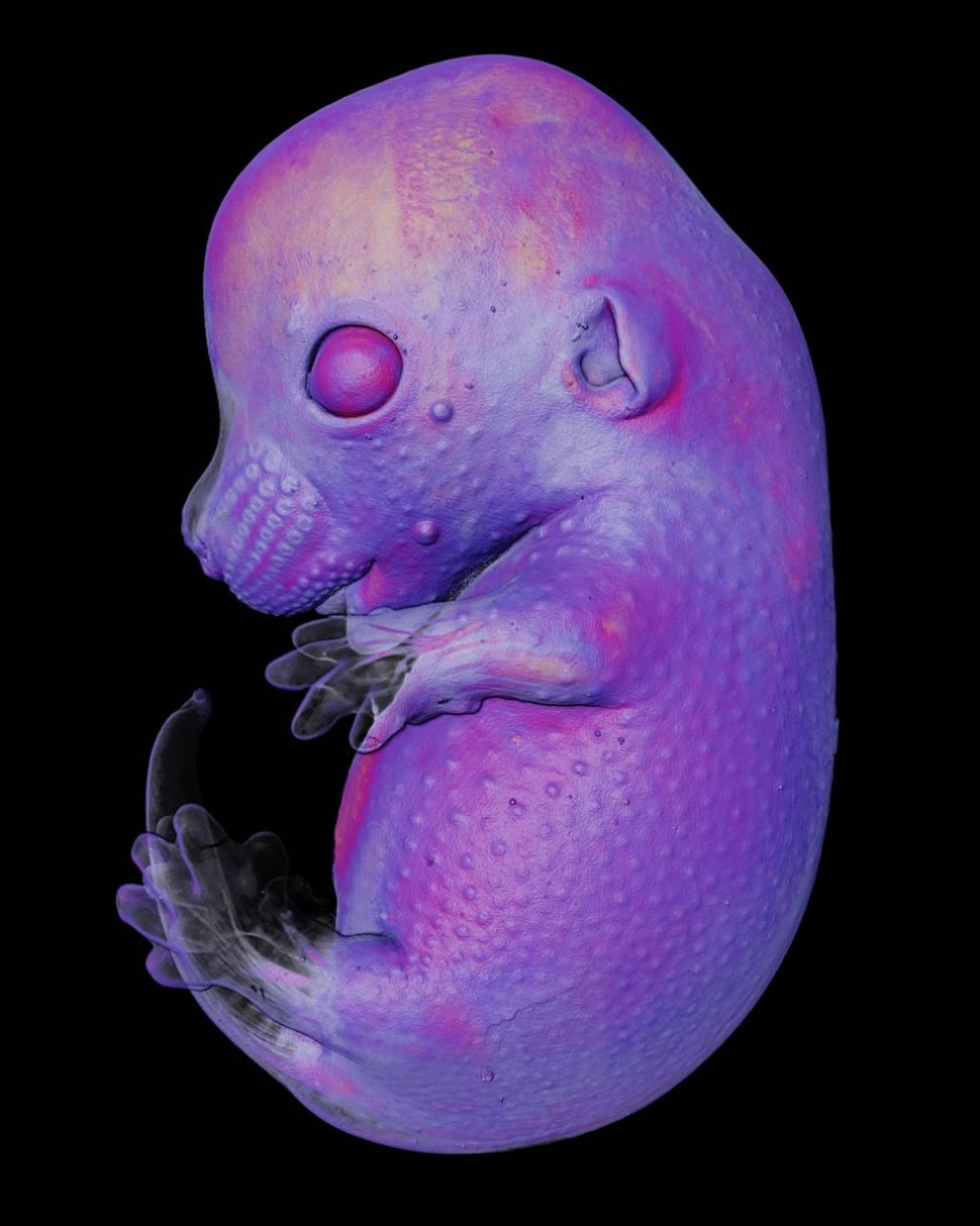
Mouse embryo by Dr. Grigorii Timin & Dr. Michel Milinkovitch
Caffeine crystals by Stefan Eberhard
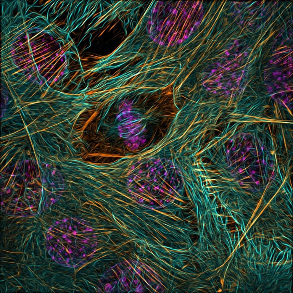
Cytoskeleton of a dividing myoblast; tubulin (cyan), F-actin (orange) and nucleus (magenta) by Vaibhav Deshmukh
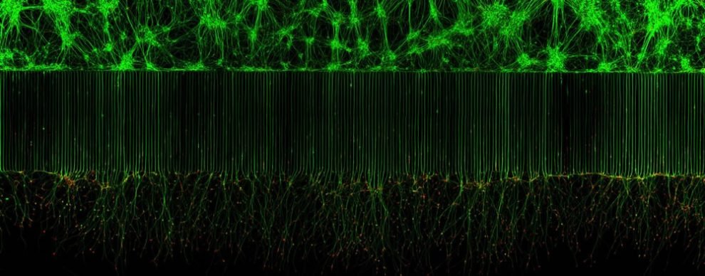
Motor neurons grown in microfluidic device for separation of cell bodies (top) and axons (bottom). Green – microtubules; Red – growth cones (actin) by Melinda Beccari & Dr. Don W. Cleveland
Crystallized sugar syrup by Dr. Diego García
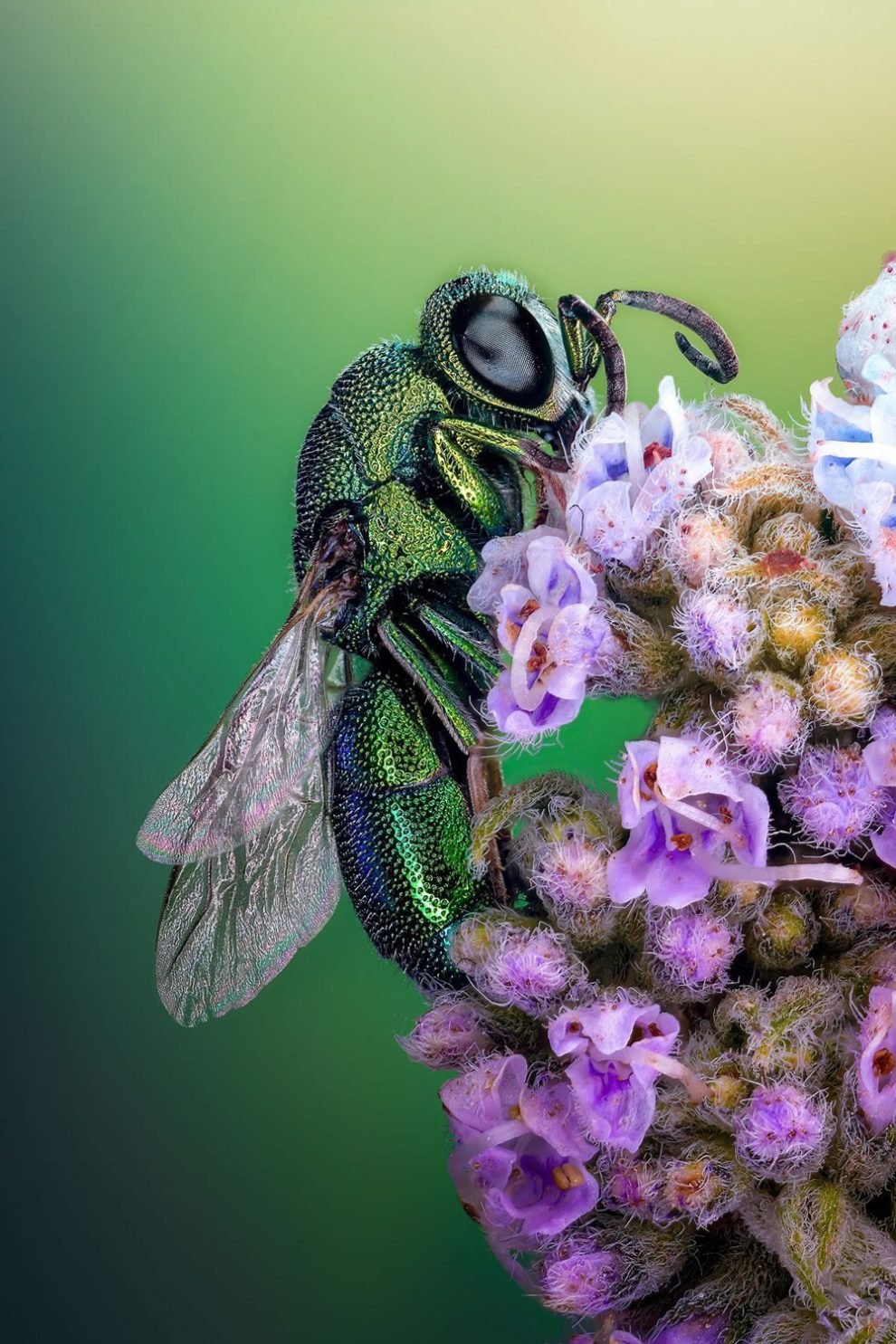
Cuckoo wasp standing on a flower by Sherif Abdallah Ahmed
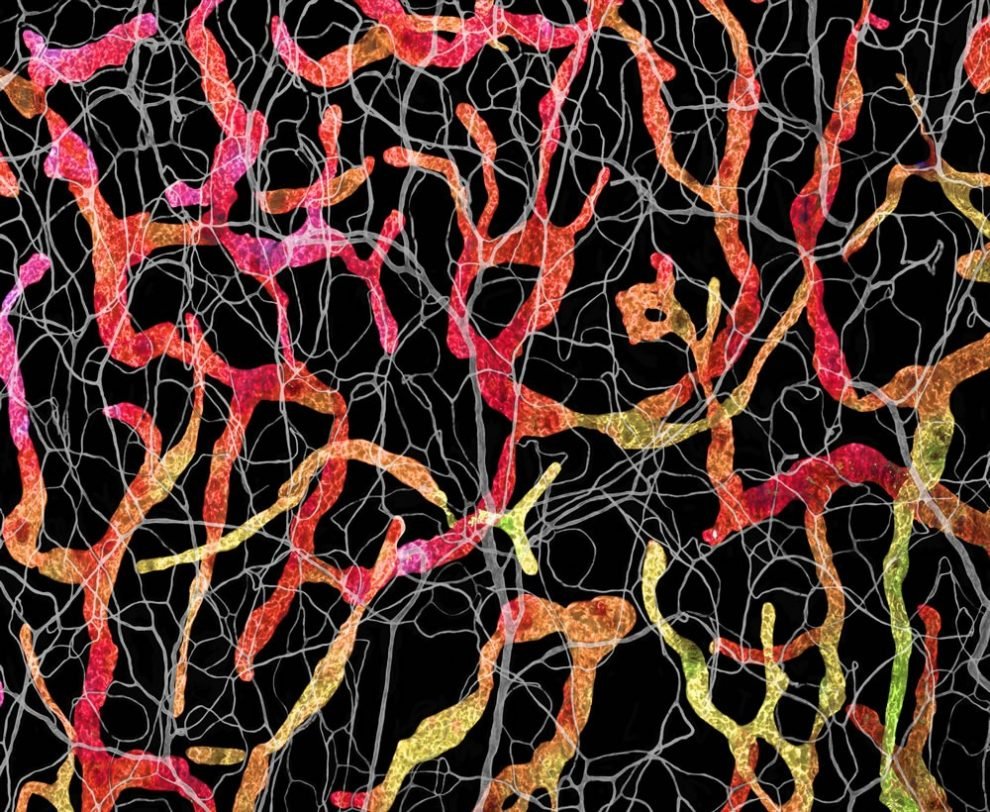
Blood and lymphatic vasculatures in the ear skin of an adult mouse by Satu Paavonsalo & Dr. Sinem Karaman
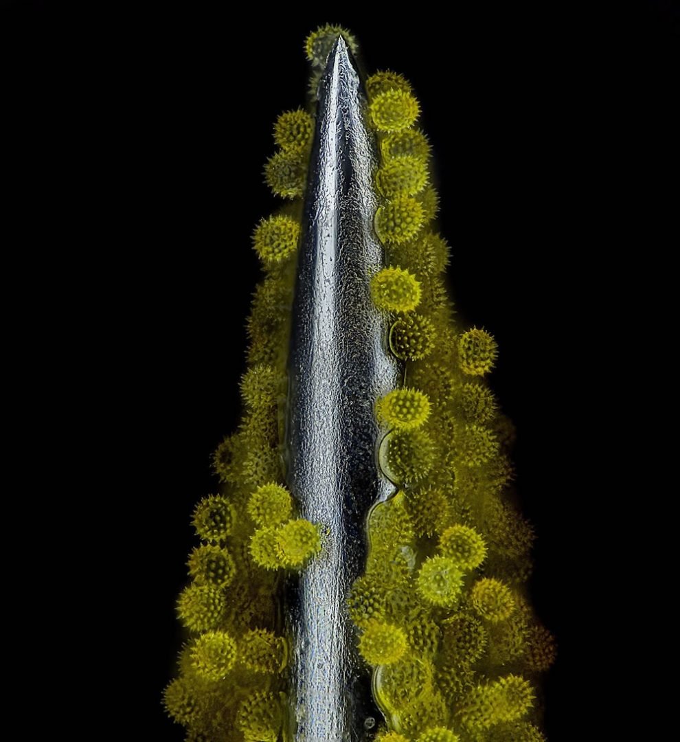
Sunflower pollen on an acupuncture needle by John-Oliver Dum
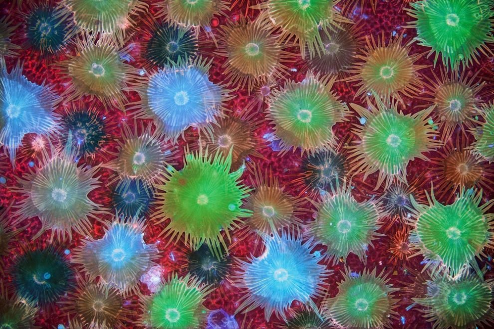
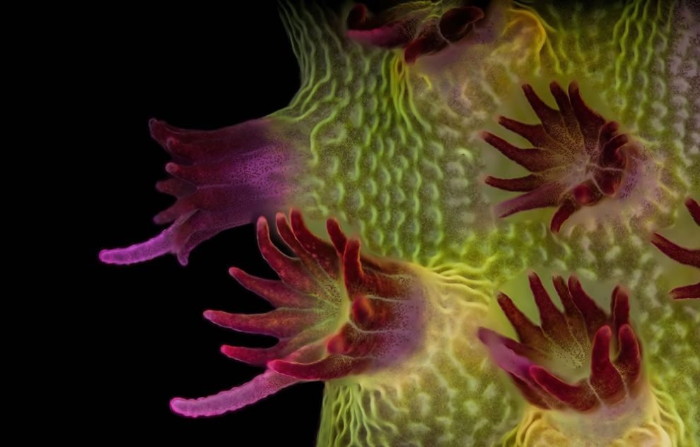
Fluorescent image of an Acropora sp. showing individual polyps with symbiotic zooxanthellae by Dr. Pichaya Lertvilai
Carbon nanotubes by Dr. Diego García
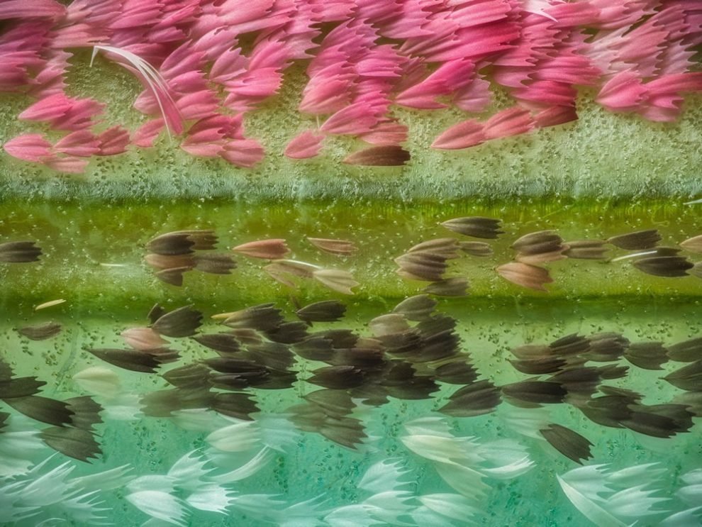
Chinese moon moth (Actias ningpoana) wing scales by Yuan Ji
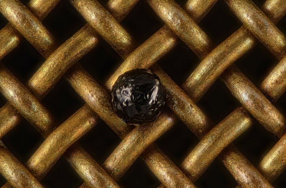
A cryptocrystalline micrometeorite resting on a #80 testing sieve by Scott Peterson
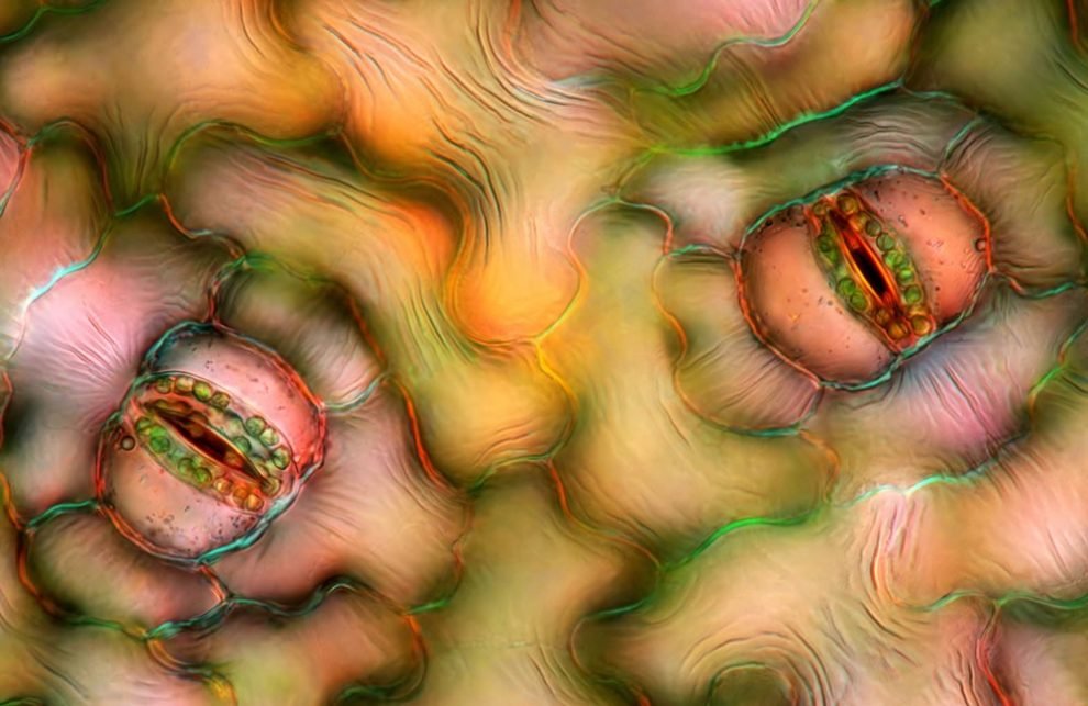
Stomata in peace lily (Spathiphyllum sp.) leaf epidermis by Marek Miś
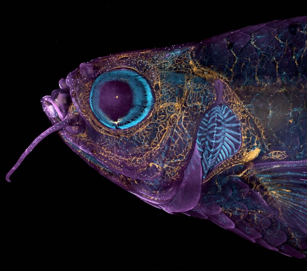
Adult transgenic zebrafish head showing blood vessels (blue), lymphatic vessels (yellow), and the skin and scales (magenta) by Daniel Castranova & Dr. Brant M. Weinstein


![[image] 170734](https://buzzbloq.com/wp-content/uploads/2024/06/1718715940_The-Superb-Tulle-Sculptures-by-Benjamin-Shine-raquo-Design-You-300x300.jpg)
![[image] 71854](https://buzzbloq.com/wp-content/uploads/2024/06/1718715692_Alison-Friend8217s-Cute-And-Funny-Animal-Drawings-Will-Completely-Melt-300x300.jpg)